Cartiliage Reconstruction (Mosaicplasty)
Mosaicplasty of the Talus
This procedure is also known as OATS – Osteoarticular transplants.
This operation is for isolated cartilage defects of the ankle (talus or tibia) that have failed treatment by 'keyhole' surgery or arthroscopy. The technique involves taking a cylinder of healthy cartilage and bone from the knee and implanting it into the ankle defect (usually the talus), which has been already been cored out. The surgery to harvest the knee cartilage is usually performed arthroscopically. The access to the ankle joint defect is usually very restricted and requires fracturing one of the bones of the ankle (medial or lateral malleolus), to expose the cartilage lesion. At the end of the procedure the ankle fracture is repaired, using standard ankle trauma techniques, with 2 screws.

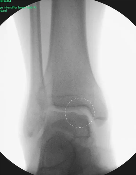
Pre operative xray showing defect in the talar cartilage.

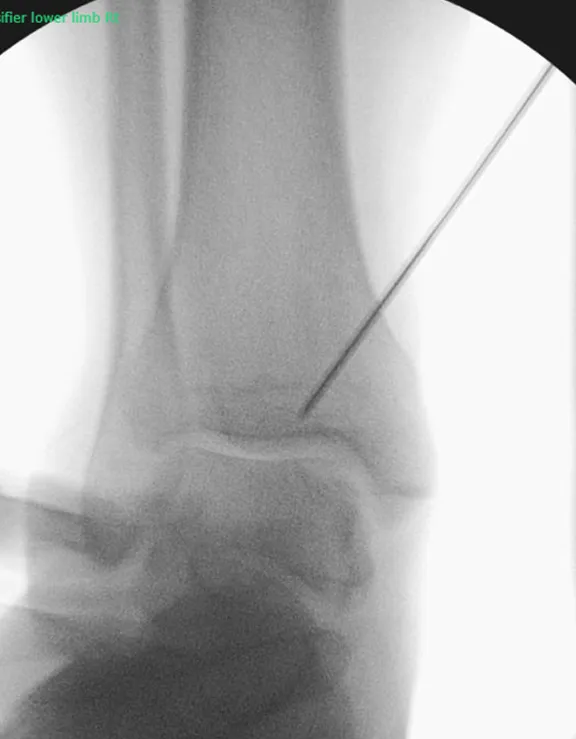
Intra operative image showing a guide wire to direct the cut in the tibia togain exposure of the talar defect. It’s important this cut is made deep enough to expose the whole defect - this stepis performed using ankle arthroscopy to ensure that the guide wire is position correctly. Once in place a saw is used to cut over the guide wire and the piece of tibia is then folded down out of the way.
The cartilage defect is then drilled out and a perfectly matching piece of healthy cartilage taken from the knee joint.

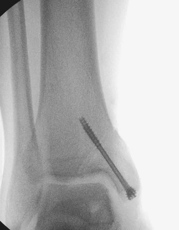
The “break” in the ankle is then fixed back together with screws.
Elevation of the foot (above the pelvis) for the first 14 days is vitally important to prevent infection. Naturally, small periods of walking and standing are necessary, but no weight must be taken through this leg for 6 weeks.
After 2 weeks the backslab will be removed and the stitches taken out, here in clinic. Another non-weightbearing plaster is applied for a further 4 weeks. At this stage you will be reviewed in clinic with xrays.
Risks of surgery
Swelling & Stiffness
Initially the ankle will be very swollen and needs elevating. The swelling will disperse over the following weeks & months but will still be apparent at 6-9 months. The ankle will be stiff when it comes out of plaster and usually requires phyiotherapy treatment.
Infection
This is the biggest risk with this type of surgery - Intravenous antibiotics are given during surgery to prevent against it. The best way to prevent an infection occurring is to keep the foot well elevated for 14 days. If there is a mild infection, it often resolves with oral antibiotics. If the infection is severe, it may warrant admission to hospital and intravenous antibiotics. A severe infection often results in failure of the procedure, and extremely rarely may result in an amputation.
Nerve Damage
The top of the foot has 3 nerves – the superficial peroneal, deep peroneal and saphenous nerves. They supply sensation to the top and sides of the foot and toes. If damaged during surgery, this will cause a numb area - which may be temporary or permanent. There is approximately a 10-15% of this happening.
Activity and time off work
- Up to 4 weeks off work is required for sedentary posts
- 8 weeks for standing or walking posts
- 12 weeks for manual / labour intensive posts
We will provide a sick note for the first 2 weeks, further notes can be obtained from the GP.
Recovery from surgery
Elevation of the foot (above the pelvis) for the first 14 days is vitally important to prevent infection. Naturally, small periods of walking and standing are necessary, but no weight must be taken through this leg for 6 weeks.
After 2 weeks the backslab will be removed and the stitches taken out, here in clinic. Another non-weight bearing plaster is applied for a further 4 weeks. At this stage you will be reviewed in clinic with x-rays.